Histamine intolerance – gut microbiome composition offers a new perspective on the issue
Histamine intolerance (HIT) is a relatively modern diagnosis that is often accompanied by questionable or incorrect diagnostic practices. This article is written to provide a clearer overview and a comprehensive picture of this condition.

Histamine intolerance (HIT) is a relatively modern diagnosis that is often accompanied by questionable and incorrect diagnosis. This article is written to provide a better overview and a comprehensive picture of this diagnosis. In the introduction, the knowledge about biogenic amines, histamine biochemistry and its degradation in the body is summarized and then HIT is summarized in terms of the cause of the disease, diagnosis and treatment.
1. Biogenic amines
Biogenic amines (BAs) are low molecular weight substances that are formed by decarboxylation of free amino acids. The main actor (biocatalyst) in the BA formation reaction is the enzyme decarboxylase, which is commonly found in various species of bacteria (both G+ and G- bacteria). However, BAs can also be formed via reductive amination and transamination of aldehydes and ketones or are formed as a result of body tissue activity. In general, they have an indispensable position in cellular metabolic activity whether in microorganisms, plants, animals or humans, in which BAs are known to have many important physiological functions. Among the many, they are involved in the synthesis of proteins, hormones and nucleic acids, promoting normal cell growth and proliferation, maintaining proper blood pressure and body temperature, influencing membrane stability, and most BAs also act as neurotransmitters, i.e., transmitters of nerve excitement [1,2].
Thus, their presence is very important in the human body however, their concentration in the body exceeding a safe limit causes many problems associated with headaches, cold sweat, gastrointestinal problems, pseudoallergic reactions and many others. The way in which biogenic amines enter the body and increase the concentration of BAs above the safe limit is primarily through the consumption of foods high in BAs, which are mainly high-protein foods (meat and meat products, fish) and foods that have undergone a fermentation process (wine, beer, soy sauce) or long-ripened foods (cheese). However, fruit, vegetables, chocolate, eggs or dairy products also contain BA. Increased BA content is also found in foods of low hygienic quality where BAs are considered as an indicator of microbial activity, since, as mentioned above, their synthesis is directly linked to the presence of microorganisms (and the decarboxylases they produce) [1,2]. Since the synthesis of biogenic amines is directly linked to the presence of microorganisms, another possible reason for their elevated concentration in the body is the improper composition of the gut microbiota and the predominance of microorganisms that tend to produce BAs in higher amounts, or the imbalanced state of the gut microbiota and the production of bacterial metabolites that may adversely affect human health.
1. 1 Histamine
Histamine is a biogenic amine that is synthesized by an enzymatic reaction - decarboxylation from the histamine precursor, histidine. This reaction was first described by Windaus and Vogt in 1907 and involves the enzyme L-histidine decarboxylase, which requires vitamin B6 (pyridoxal phosphate) as a cofactor. The physiological and pathophysiological effects of histamine were first described in 1910 by Dahl and Laidlaw, who are considered pioneers in histamine research. In 1932, histamine was identified as a mediator of anaphylactic reactions [3,4].
Today, many effects of histamine in the human body are now known. Histamine is synthesized by basophils, mast cells, platelets, histaminergic neurons, and enterochromaffin cells of the gastrointestinal tract, where histamine is stored intracellularly in vesicles and is released upon stimulation. In addition, other cell types such as neutrophils and lymphocytes have the ability to synthesize histamine de novo. In these cases, we speak of endogenous histamine. In general, histamine is involved in various immunological and physiological mechanisms through the binding of histamine to its four receptors (H1, H2, H3, and H4) located on target cells in various tissues. The aforementioned mediated immunological and physiological mechanisms include stimulation of gastric acid secretion, inflammation, smooth muscle cell contraction, vasodilation, increased vascular permeability and mucus secretion, tachycardia, changes in blood pressure, arrhythmia, cytokine production, and various others. In addition, histamine is known for many other functions in neurotransmission, immunomodulation, hematopoiesis, wound healing, day-night rhythms, regulation of histamine- and polyamine-induced cell proliferation, and angiogenesis in tumor models and intestinal ischemia. This diversity and the large spectrum of nonspecific gastrointestinal and extraintestinal symptoms is due to the wide distribution of the four histamine receptors in different organs and tissues in the body [4-6].
1.1.1 Histamine clearance in the body
Two major metabolic pathways involving either the enzyme diamine oxidase (DAO) or histamine-N-methyltransferase (HNMT) serve to degrade histamine in the body. These two enzymes are functionally and structurally distinct from each other and are involved in histamine degradation in different ways(Fig.1).
DAO catalyzes the oxidative deamination reaction of the primary amino group on the histamine molecule. It is a secretory protein that is stored in the plasma membrane in vesicular structures in epithelial cells and is secreted into the circulation at a certain stimulus and is responsible for the degradation of extracellular histamine. In animals, DAO expression occurs only in certain tissues such as the small intestine, ascending colon, placenta, and kidney. In the intestine, DAO activity increases from the duodenum to the ileum and is mainly localized in intestinal villi [4,5].
In contrast, HNMT is expressed in almost all tissues in the kidney, liver, spleen, colon, prostate, ovary, spinal cord cells, trachea and respiratory tract. HNMT catalyzes the methylation of the secondary amino group on the histamine imidazole and, unlike DAO, is a cytosolic enzyme that is responsible for the metabolism of intracellular histamine. The two enzymes also differ in substrate specificity. HNMT is highly selective for histamine whereas DAO can also metabolize other biogenic amines such as putrescine, cadaverine although it has the highest preference for histamine. This will be mentioned in a later chapter in which the anti-histamine diet will be described [4,5].
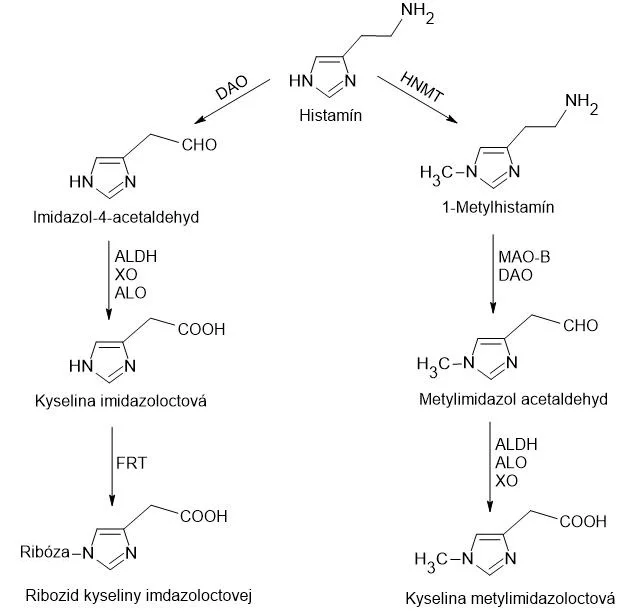
Fig.1: The main metabolic pathways of histamine. In the human body, histamine metabolism can be twofold:
1. nitrogen in the imidazole cycle is methylated by HNMT to form N-methylhistamine, which is subsequently oxidized by MAO-B or DAO to methylimidazole acetaldehyde, and oxidized to methylimidazoleacetic acid by the action of ALDH, ALO or XO.
2. The action of DAO produces imidazole-4-acetaldehyde, which is subsequently oxidized to imidazoleacetic acid by ALDH, XO or ALO, which binds to ribose via FRT. DAO: diamine oxidase; ALDH: aldehyde dehydrogenase; XO: xanthine oxidase; ALO: aldehyde oxidase; FRT: phosphoribosyltransferase; HNMT: histamine-N-methyltransferase; MAO-B: monoamine aminobutyric acid oxidase type B.
Depending on the localization, we know endogenous and exogenous histamine and in the case of HIT it is the exogenous histamine that is of interest and therefore will be the most discussed in this thesis. Exogenous histamine is ingested through food and the gateway for this histamine is the intestinal epithelium and therefore, although HNMT is also present in the gastrointestinal tract, the more highly expressed DAO plays a major role in protecting the body against exogenous histamine, which is either derived from ingested food or produced by the gut microbiota. To confirm this effect, many studies have been conducted where research teams selectively inhibit DAO and then administer doses of histamine to animal experimental models and observe this effect. HNMT is also involved to a small extent in the degradation of exogenous histamine, but appears to be more effective in degrading intravenous and intradermal histamine [5].
2. Histamine intolerance
Histamine intolerance is also commonly referred to as "enteral histaminosis". This disease is caused by a deficiency of the gastrointestinal enzyme DAO and thus an inability to metabolize histamine, with the lack of DAO activity being due to either a congenital defect (polymorphisms - SNPs ingene encoding DAO), inhibition of this enzyme by specific agents and drugs or due to various gastrointestinal diseases such as non-specific intestinal inflammation, dysmicrobia, etc [7,8]. With polymorphism, it is important to note that different variations in the gene encoding DAO are associated with DAO levels but not enough to cause the associated disease state of HIT. This requires the interaction of several environmental factors such as modification in the alternative N-methyltransferase pathway, vesicular transport of both enzymes and amines, or in the ability of enterocytes to absorb histamine [9]. HIT is always an adverse reaction to foods without immunological basis, which distinguishes this disease from classical food allergy. According to the World Allergy Organization, food intolerance, i.e. an adverse reaction to food without an immunological basis, should be referred to as non-allergic food hypersensitivity to clearly distinguish it from food allergy, which is invariably triggered by a specific immune mechanism. Thus, histamine intolerance is a disorder that arises due to a reduced ability to degrade histamine in the gut due to impaired DAO activity, leading to the accumulation of histamine in the plasma and the occurrence of adverse effects [7].
Although the first scientific references to histamine intolerance date back more than 20 years, it is only in the last decade that there has been a significant increase in research interest in the subject, as reflected in the following graph, which was created based on the results of a search of the MEDLINE bibliographic database (PubMed, Fig. 2).

The manifestations of histamine intolerance vary precisely because of the wide distribution of the four histamine receptors in different organs and tissues of the body(Fig.3). In the work of the research team of Schnedl et al. followed the symptoms of 133 patients who suffered from gastric discomfort and in whom diseases that could manifest similar symptoms to HIT, namely lactose intolerance, fructose malabsorption, H. pylori infection and celiac disease, were excluded by the available methods. Among all manifestations, gastrointestinal manifestations such as abdominal distension (92%), postprandial fullness, diarrhea, abdominal pain, and constipation (55-73%) were the most common. Gastrointestinal manifestations were followed by neurological and cardiovascular manifestations such as dizziness, headache, palpitations and others (47-66%) and finally respiratory and dermatological symptoms, which accounted for 26-48% of all symptoms reported. Up to 97% of patients reported more than 3 symptoms simultaneously after receiving a higher dose of histamine with a median number of symptoms of 10, indicating a high correlation between histamine doses and symptom occurrence [10]. This high number of different symptoms and manifestations that are not strictly disease-specific complicates the diagnosis of HIT and contributes to the difficulty in establishing uniformity about the diagnostic criteria for HIT.
![Hlavné príznaky HIT a pravdepodobne zodpovedajúce histamínové receptory. Prebrané z [5]](https://iprobio.venalio.com/media/2025/05/0/1/histaminova-intolerancia-5-size-banner-large-v-2.webp)
Fig.3: Main symptoms of HIT and probably corresponding histamine receptors. Adapted from [5].
2.1 Diagnosis of HIT
Although significant progress has been made in the elucidation of histamine intolerance in the last decade, the diagnosis of this disease remains a challenge precisely because of the non-specificity of symptoms and the lack of validated diagnostic tools. Despite the difficulty of diagnosis, Basté et al. in their work proposed a schematic summary of the diagnostic algorithm based on the current scientific knowledge(Fig. 4). According to authors dealing with the diagnosis of HIT, it is necessary and important to first exclude other potential causes of symptoms associated with elevated plasma histamine levels [5,8,11,12]. For this purpose, skin prick tests, also called "skin prick tests", are used and recommended to exclude IgE sensitization caused by food allergy. It is also important to know whether the patient is taking medications that inhibit DAO activity. If these conditions are negative and at least two typical symptoms of HIT are confirmed and there is an improvement in health after adherence to an antihistamine diet, the diagnosis of HIT will be confirmed [5].
![Zhrnutie popísaného prístupu k diagnostike HIT. Prebrané z [5]](https://iprobio.venalio.com/media/2025/05/0/1/histaminova-intolerancia-6-size-banner-large-v-2.webp)
Fig.4: Summary of the described approach to the diagnosis of HIT. Taken from [5].
2.2 Approaches to the treatment of HIT
Currently, one of the main approaches leading to the alleviation and prevention of histamine intolerance symptoms is the adherence to a low histamine diet. Another approach is exogenous supplementation of DAO with appropriate nutritional supplements. Very often, a combination of these two approaches is used to effectively alleviate and suppress the symptoms of HIT [5].
2.2.1 Low-histamine diet
As previously mentioned, a low-histamine diet has so far been considered the main strategy in the preventive treatment of HIT. This diet excludes different types of foods that may contain high levels of histamine, however, there is no clear dietary recommendation and it is necessary to monitor food sensitivities and histamine content on an individual patient basis with regard to foods containing higher histamine levels [5]. The following table represents the foods that are excluded from the various low-histamine diets found in the literature(Table 1).
Tab.1: Excluded foods in various low-histamine diets found in the literature. Adapted from [5].
Excluded foods from low-histamine diets | ||
<20* | 20-60%* | >60%* |
Milk | Shellfish | Ripe and semi-ripened cheeses |
Lentils | Eggs | Grated cheeses |
Chickpeas | Fermented soya products | Oily fish |
Soya beans | Eggplant | Canned oily fish |
Mushrooms | Avocado | Dry-fermented meat |
products | ||
Banana | Spinach | |
Kiwi | Tomatoes | |
Pineapple | Sauerkraut | |
Plum | Citrus | |
Nuts | Strawberries | |
Chocolate | Wine | |
Beer | ||
* Percentage of low-histamine diets from the literature that exclude each food.
Excluding the foods in Table 1 however, HIT patients must also beware of foods that contain other biogenic amines such as putrescine or cadaverine, since, as mentioned above, the DAO enzyme is not strictly specific for histamine, but can also metabolize other amines (acting as competitive substrates) thus slowing down the metabolism of histamine andincreases its plasma level if other biogenic amines are present in significant amounts. Examples of such foods are citrus fruits, mushrooms, soybeans, bananas and nuts. In addition, some diets exclude other foods that do not contain histamine or other amines that would justify the exclusion of these foods, but are reported to cause the release of endogenous histamine, although the corresponding mechanism has not yet been elucidated. Examples of such foods are papaya, kiwifruit, strawberries, pineapple, and plums [5].
2.2.2 Exogenous DAO supplements
A treatment similar to that for lactose intolerance, namely oral supplementation of an enzyme, in this case DAO, has been proposed to improve the breakdown of dietary histamine. This would also allow patients to follow a less restrictive diet, including a diet with a tolerable dose of histamine. However, the problem is that there are currently only five published intervention studies that have tested the clinical efficacy of exogenous DAO supplementation in patients with HIT symptoms. Although the results of these studies are promising, and in all of them there was either a reduction in the intensity and frequency of symptoms [13-15], an increase in plasma DAO activity (in 61% of the patients tested) [15] or a reduction in the severity of chronic spontaneous urticaria andmigraine, which are diseases associated with HIT [16,17], further clinical trials with a larger patient sample and a rigorous experimental design are needed to unequivocally confirm the efficacy of this treatment [5].
Regarding the DAO supplement itself, in 2017, the European Commission authorized the marketing of DAO supplement as a dietary supplement or as a food for special medical purposes. These European regulations allow the DAO supplement in the form of an enteric-coated porcine kidney protein extract to ensure integrity during passage through the gastric environment [5].
Porcine kidney is generally considered to be a major source of DAO enzyme with a demonstrated ability to degrade histamine and other biogenic amines in vitro [18-22], however, some research teams have demonstrated a higher catalytic capacity of plant-derived DAO enzymes[23,24], which may be of commercial interest to the vegetarian/vegan population, but also to people with religious restrictions on the consumption of pork products. In particular, germinated sprouts of certain edible legumes are interesting sources of plant-derived DAOs [ 25]. Germination is a physiological process in which the enzymatic capacity of DAO can increase up to 250-fold compared to ungerminated seeds, and the increased presence of DAO enzyme in legume sprouts is thought to be related to the importance of hydrogen peroxide in cell wall structuring, lignification, and mobilization of seed reserves during germination [5,26-28].
In addition to these two main strategies in the treatment of HIT, it is important to think of other approaches that can alleviate the symptoms of HIT and go after the cause of the disease. It is possible to experiment with supplements that are precursors of endogenous DAO production such as vitamin B6 (in the P-5-P enzyme), copper, and vitamin B2. In addition, vitamin C may be helpful in breaking down excess histamine [ 29]. However, there is increasing emphasis on the gut microbiome and the importance of its treatment in association with HIT. Therefore, the following chapter will be devoted to this context.
2.3 HIT and the gut microbiome
Associations between various diseases and the gut microbiome are increasingly emerging and this trend is expected to continue. This is no different in the case of HIT. However, only one study has been published so far that has looked at differences in the composition of the gut microbiome in healthy patients, HIT patients and patients suffering from other food intolerances or allergies. Although only 64 participants took part in this study, the results are very interesting.
In the study by Schink et al. 64 participants were followed up, with 33 patients having suspected HIT. Further profiling and measurement of DAO activity in these patients revealed 8 of the 33 patients with HIT by definition. The remaining 25 patients, with normal DAO activity, were considered food hypersensitive. Furthermore, 21 patients with proven food allergy and 10 healthy volunteers without gastrointestinal discomfort participated in this study [30].
This study found several facts and interesting features in the composition of the microbiome between these groups. The research team investigated several families and their individual representatives. In the case of the Enterobacteriaceae family, there were no significant differences between the groups, although there was a slight increase of Enterobacteriaceae by 0.17% in the HIT patients. However, when focusing on the Proteobacteria strain, which belongs to the Enterobacteriaceae family, differences were observed in the HIT patients, namely an increased representation of this strain among the other strains belonging to this family compared to the healthy group. According to some studies, the increased abundance of Proteobacteria may indicate intestinal dysbiosis and/or altered epithelial function in this group of patients [30-33].
Bacterial strains within the Lactobacillus family, e.g. Lactobacillus casei or Lactobacillus delbrueckii, show histidine decarboxylase activity, which is involved in histamine production. Interestingly, however, none of the participants in the study showed increased levels of Lactobacillus bacteria and therefore the influence of bacterial histamine as a cause of increased intestinal histamine exposure in histamine-intolerant individuals is questionable. A more thorough study and a more detailed characterization of the bacteria at the species level is needed for clarification. However, dysbiosis may promote mucosal inflammation in the gut, and since the DAO enzyme is synthesized by enterocytes and stored in mucosal epithelial cells, disruption of these cells caused by inflammation may lead to decreased DAO synthesis. This may further lead to decreased degradation of exogenous histamine and to increased levels of endogenous histamine, which causes the typical symptoms of HIT. The research team of Schink et al. however, did not study the association between dysbiosis and intestinal inflammation, although these studies are prospective for the future.
Instead, they observed a significantly higher abundance of the bacterial genus Roseburia in patients suffering from HIT compared to healthy controls and other groups. The bacterial genus Roseburia produces butyrate by default, and various health-promoting effects have been attributed to this bacterial genus. It has even been found that patients suffering from chronic kidney disease or those with ulcerative colitis have reduced amounts of Roseburia sp. In a study by Schink et al. however, patients with HIT showed an increased proportion of Roseburia. They justify this fact by the fact that bacteria of the genus Roseburia produce short-chain fatty acids in the colon from indigestible carbohydrates such as starch, inulin, or xylan, and thus these substrates promote their metabolism andtherefore, the increased proportion of this genus may be due to the patients' diet, which is rich in carbohydrates and fibre, which may have prebiotic properties and promote the growth of butyrate-producing bacteria [30].
However, patients with HIT showed significantly lower levels of the bacterial genus Butyricimonas, which is also responsible for butyrate production. In this case, reduced butyrate levels in the colon may lead to impaired barrier function and the development of inflammatory bowel disease. It may be that fewer of these bacteria may have adverse effects on the health of patients with HIT. Although the effects of butyrate are beneficial and play an important role as an energy source for enterocytes, on the other hand, there are also contrary reports in the case of excess butyrate and its adverse effects on human health [30].
The Bifidobacteriaceae family contains many bacterial species beneficial to human health and the importance of the presence of bacterial species from this family in the colon is well known to the general public, especially due to the increasing number of advertisements targeting various food products containing these beneficial bacteria. In a study by Schink et al. a relationship was observed between healthy patients and increased abundance of bacteria from the Bifidobacteriaceae family (called bifidobacteria). The essence of the beneficial effect of these bacteria lies in the lowering of intestinal pH by the production of acetic and lactic acid, which limits the growth of pathogenic bacteria and blocks adhesion to the intestinal mucosa. Decreased numbers of bifidobacteria have been observed in several disorders including allergies, irritable bowel syndrome and inflammatory bowel disease.
Further, a study by Schink et al. verrucomicrobia, which was only elevated in the food hypersensitivity group, but on the contrary, in the HIT patients the abundance of this bacterial strain was very low. In relation to this strain, high colonic colonisation has been described in patients treated with broad-spectrum antibiotic therapy.
The results of the work of Schink et al. suggest a change in microbial composition in food intolerances, especially in patients with histamine intolerance. Increased abundance of bacteria from the Proteobacteria family, decreased abundance of Bifidobacteriaceae/Bifidobacterium species and lower bacterial diversity point to dysbiosis and a disrupted intestinal barrier in this group of patients. The positive correlation between stool histamine and zonulin levels suggests a negative effect of histamine on intestinal permeability. However, the authors of the study did not find increased histamine concentrations in stool samples of patients with HIT or increased abundance of histamine-producing bacteria; nevertheless, dysbiosis in patients may contribute to mucosal inflammation, which may lead to decreased synthesis of the DAO enzyme and consequently increased histamine levels and clinical symptoms in susceptible patients. Future studies with larger numbers of participants and well-designed experimental parameters, such as the identification of mucosal-bound bacteria instead of stool bacteria, are needed to confirm these preliminary findings.
3. Conclusion
Although histamine intolerance is a frequently discussed topic, there are still many unanswered questions regarding this disease. However, as can be seen from the graph in Fig. 2 in Ch. 2, interest in this topic is increasing as new facts and connections are emerging that have not yet been elucidated and that provide new insights into the issue of HIT. One of these is the link between the composition of the gut microbiome and HIT, which is becoming increasingly important. Although there are still many question marks on this topic precisely because of the lack of experimental studies aimed at identifying the gut microbiome in detail in HIT patients, there is a growing awareness of the enormous impact of the gut microbiome on host immunological processes. In this case, not only the taxonomic diversity of bacteria plays a major role, but also bacterial metabolites, which also have an important impact on human health.
Author. Mária Bláhová, Department of Biotechnology, Faculty of Chemical and Food Technology, STU in Bratislava
4. Literature:
1.wójcik, W., Łukasiewicz, M. & Puppel, K. Biogenic amines: formation, action and toxicity-a review. (2020) doi:10.1002/jsfa.10928.
2.kohajdová, Z. & Karovičová, J. Biogenic amines - formation, methods of determination and occurrence in food. 79-85 (2001).
3.dale, H. H. & Laidlaw, P. P. The physiological action of β-iminazolylethylamine. J. Physiol. 41, 318-344 (1910).
4.maintz, L. & Novak, N. Histamine and histamine intolerance. Am. J. Clin. Nutr. 85, 1185-1196 (2007).
5.comas-Basté, O., Sánchez-Pérez, S., Veciana-Nogués, M. T., Latorre-Moratalla, M. & Vidal-Carou, M. D. C. Histamine intolerance: the current state of the art. Biomolecules 10, 1-26 (2020).
6.jutel, M., Akdis, M. & Akdis, C. A. Histamine, histamine receptors and their role in immune pathology. Clin. Exp. Allergy 39, 1786-1800 (2009).
7.schnedl, W. J. & Enko, D. Histamine intolerance originates in the gut. Nutrients 13, 1-14 (2021).
8.hanuskova, E. & Plevková, J. Histamine intolerance Histamine intolerance. Cesk. Physiol. 62, 26-33 (2013).
9.maintz, L. et al. Association of single nucleotide polymorphisms in the diamine oxidase gene with serum diamine oxidase activities. Allergy Eur. J. Allergy Clin. Immunol. 66, 893-902 (2011).
10.schnedl, W. J. et al. Evaluation of symptoms and symptom combinations in histamine intolerance. Intest. Res. 17, 427-433 (2019).
11.tuck, C. J., Biesiekierski, J. R., Schmid-Grendelmeier, P. & Pohl, D. Food Intolerances. doi:10.3390/nu11071684.
12.comas-Basté, O., Latorre-Moratalla, M. L., Bernacchia, R., Veciana-Nogués, M. T.. & Vidal-Carou, M. C. New approach for the diagnosis of histamine intolerance based on the determination of histamine and methylhistamine in urine. J. Pharm. Biomed. Anal. 145, 379-385 (2017).
13.manzotti, G., Breda, D., Di Gioacchino, M. & Burastero, S. E. Serum diamine oxidase activity in patients with histamine intolerance. Int. J. Immunopathol. Pharmacol. 29, 105-111 (2016).
14.komericki, P. et al. Histamine intolerance: Lack of reproducibility of single symptoms by oral provocation with histamine: A randomized, double-blind, placebo-controlled cross-over study. Vienna. Klin. Wochenschr. 123, 15-20 (2011).
15.schnedl, W. J. et al. Diamine oxidase supplementation improves symptoms in patients with histamine intolerance. Food Sci. Biotechnol. 28, 1779-1784 (2019).
16.izquierdo-Casas, J. et al. Diamine oxidase (DAO) supplementation reduces headache in episodic migraine patients with DAO deficiency: A randomized double-blind trial. Clin. Nutr. 38, 152-158 (2019).
17.yacoub, M. R. et al. Diamine Oxidase Supplementation in Chronic Spontaneous Urticaria: A Randomized, Double-Blind Placebo-Controlled Study. Int. Arch. Allergy Immunol. 176, 268-271 (2018).
18.comas-Basté, O., Latorre-Moratalla, M. L., Sánchez-Pérez, S., Veciana-Nogués, M. T.. & Vidal-Carou, M. C. In vitro determination of diamine oxidase activity in food matrices by an enzymatic assay coupled to UHPLC-FL. Anal. Bioanal. Chem. 411, 7595-7602 (2019).
19.kettner, L., Seitl, I. & Fischer, L. Evaluation of porcine diamine oxidase for the conversion of histamine in food-relevant amounts. J. Food Sci. 85, 843-852 (2020).
20.mondovì, B. et al. Diamine oxidase from pig kidney. Improved purification and properties. J. Biol. Chem. 242, 1160-1167 (1967).
21.floris, G., Fadda, M. B., Pellegrini, M., Corda, M. & Agro', A. F. Purification of pig kidney diamine oxidase by gel-exclusion chromatography. FEBS Lett. 72, 179-181 (1976).
22.bouvrette, P., Male, K. B., Luong, J.. H. T. & Gibbs, B. F. Amperometric biosensor for diamine using diamine oxidase purified from porcine kidney. Enzyme Microb. Technol. 20, 32-38 (1997).
23.pietrangeli, P., Federico, R., Mondovì, B. & Morpurgo, L. Substrate specificity of copper-containing plant amine oxidases. J. Inorg. Biochem. 101, 997-1004 (2007).
24.masini, E. et al. Pea seedling histaminase as a novel therapeutic approach to anaphylactic and inflammatory disorders: A plant histaminase in allergic asthma and ischemic shock. ScientificWorldJournal. 7, 888-902 (2007).
25.comas-Basté, O., Latorre-Moratalla, M. L., Rabell-González, J., Veciana-Nogués, M. T.. & Vidal-Carou, M. C. Lyophilised legume sprouts as a functional ingredient for diamine oxidase enzyme supplementation in histamine intolerance. Lwt 125, 109201 (2020).
26.joseph, P. & Srivastava, S. K. Photoregulation of Diamine Oxidase from Pea Seedlings. J. Plant Physiol. 146, 108-114 (1995).
27.laurenzi, M. et al. Analysis of the distribution of copper amine oxidase in cell walls of legume seedlings. Planta 214, 37-45 (2001).
28.tavladoraki, P., Cona, A. & Angelini, R. Copper-containing amine oxidases and FAD-dependent polyamine oxidases are key players in plant tissue differentiation and organ development. Front. Plant Sci. 7, (2016).
29.nichols, M., Mueller D., Schuler N., Medicine with the heart. Internet resource: https://medicinewithheart.com/blog/histamine-intolerance-2/. Downloaded on August 4, 2021.
30.schink, M. et al. Microbial patterns in patients with histamine intolerance. J. Physiol. Pharmacol. 69, 579-593 (2018).
31.shin, N. R., Whon, T. W.. & Bae, J. W. Proteobacteria: Microbial signature of dysbiosis in gut microbiota. Trends Biotechnol. 33, 496-503 (2015).
32.carvalho, F. A. et al. Transient inability to manage proteobacteria promotes chronic gut inflammation in TLR5-deficient mice. Cell Host Microbe 12, 139-152 (2012).
33.litvak, Y., Byndloss, M. X., Tsolis, R. M. & Bäumler, A. J. Dysbiotic Proteobacteria expansion: a microbial signature of epithelial dysfunction. Curr. Opin. Microbiol. 39, 1-6 (2017).